Pinole Aesthetic Dentistry Creating Beautiful Smiles
There’s a lot happening in your mouth that a visual exam alone cannot reveal. Decay between teeth, infections brewing at the root tip, early bone loss from gum disease, impacted teeth that haven’t broken through the surface — none of these announce themselves on the outside. Dental imaging is what closes that gap, and digital dental X-rays in Pinole, CA have made the process faster, safer, and more precise than it’s ever been.
If you’ve ever wondered what’s actually happening when your dentist takes X-rays, or why the technology matters as much as the dentist taking them, this guide answers those questions directly. Digital radiography in dentistry has fundamentally changed how oral health is diagnosed and monitored — and knowing how it works helps you make sense of your own care.
How Digital Dental X-Rays Work
Traditional film-based X-rays required chemical processing and produced a static image that couldn’t be adjusted after the fact. Digital radiography replaces film with an electronic sensor that captures the image instantly and transmits it directly to a computer screen. The entire process takes seconds, and the resulting image can be enlarged, enhanced, and analyzed in ways that film never allowed.
The sensor detects X-ray energy and converts it into a digital signal. That signal produces a high-resolution image of your teeth, roots, surrounding bone, and soft tissue structures — all with far less radiation than traditional film. Dental X-rays at Pinole Aesthetic Dentistry are a standard part of comprehensive care, giving Dr. Sisodia and Dr. Seth a complete, real-time picture of what’s happening beneath the surface of every patient’s smile.
According to the American Dental Association, digital sensors reduce radiation exposure by up to 80% compared to traditional dental X-ray film. For patients who are cautious about radiation, this is a meaningful improvement, not just a marketing claim. The dose from a full set of digital dental X-rays is comparable to a few hours of natural background radiation from everyday environmental sources.
The Imaging Technology Available at Pinole Aesthetic Dentistry
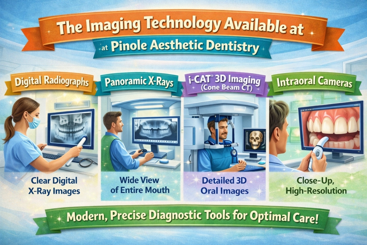
Not all dental imaging is the same. Different tools reveal different information, and a well-equipped practice uses multiple imaging modalities depending on the patient’s situation. Here’s what Pinole Aesthetic Dentistry offers and what each one does:
Digital Radiographs (Bitewing and Periapical X-Rays)
These are the most common types of X-rays taken during a routine visit. Bitewing X-rays show the upper and lower back teeth in a single image, revealing decay between teeth and any changes in bone density from gum disease. Periapical X-rays show the entire tooth from crown to root tip — useful for evaluating the root structure, detecting abscesses, and monitoring bone levels around specific teeth.
In digital form, both types produce sharper images with greater detail than film, and the sensor placement is the same as the traditional bite-tab or film packet patients are familiar with. The main difference is the speed — the image appears on the screen in seconds rather than waiting for the film to develop.
Panoramic X-Rays
Panoramic X-rays capture the entire mouth in a single wide image — all your teeth, both jaws, the temporomandibular joints (TMJ), sinuses, and surrounding bone — in one sweep. Unlike bitewings, which require multiple sensor placements, a panoramic X-ray uses a rotating arm that moves around the outside of your head.
This broad view is particularly useful for evaluating wisdom teeth, assessing jaw development, detecting cysts or tumors, and planning full-arch treatments like implants or dentures. For new patients, a panoramic X-ray is often the most efficient starting point for a comprehensive dental evaluation. Digital panoramic systems produce these large-format images in seconds with significantly lower radiation than older equipment.
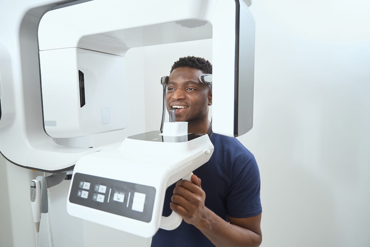
i-CAT® 3D Imaging (Cone Beam CT)
The i-CAT® 3D imaging system is a cone beam computed tomography (CBCT) scanner — a step beyond standard two-dimensional X-rays. Instead of a flat image, it produces a three-dimensional model of the jaw, teeth, nerve canals, sinuses, and surrounding anatomy that can be rotated and viewed from any angle on screen.
This level of detail is invaluable for implant planning, where precise knowledge of bone volume, density, and nerve location determines exactly where and how an implant can be placed. It’s also used for complex extractions, orthodontic assessment, and evaluating TMJ disorders. For patients considering dental implants in Pinole, this technology removes much of the guesswork from surgical planning and significantly improves outcomes.
Intraoral Cameras
While not an X-ray, an intraoral camera complements digital radiography by providing highly magnified, real-time video of the inside of your mouth from a patient’s perspective. A small, pen-sized camera captures detailed images of each tooth that are displayed on a chairside monitor — showing cracks, early decay, worn enamel, and other surface changes that a dentist’s eye alone might miss.
More importantly, intraoral cameras allow patients to see exactly what their dentists see. This transparency makes treatment discussions far more productive and helps patients make informed decisions about their care with confidence rather than uncertainty.
What Digital X-Ray Dentistry Catches That Visual Exams Miss
The clinical value of X-ray dentistry comes down to what it reveals that simply cannot be seen any other way. Interproximal cavities (decay that forms between two teeth where they contact each other) are invisible until they’re large enough to cause structural damage or pain. Digital radiographs catch these at a stage when a small composite filling is all that’s needed.
Periapical pathology (infections or cysts at the root tip) often causes no symptoms for extended periods. By the time a patient notices pain or swelling, the infection has frequently spread. A periapical X-ray identifies these lesions early, allowing treatment before surgical intervention becomes necessary.
Bone level assessment is another area where imaging is irreplaceable. Gum disease destroys the bone that supports your teeth, and that loss is clinically silent in its early stages. Regular digital radiographs track bone levels over time and detect changes that would otherwise go unnoticed until significant damage has already occurred.
See More. Know More. Treat Better.
The quality of your dental care is connected to the quality of information your dentist has. Digital dental X-rays in Pinole, CA, give Dr. Sisodia, Dr. Seth, and their team a level of diagnostic clarity that wasn’t available just a generation ago. Whether it’s a bitewing that catches a cavity before it needs a root canal, a panoramic image that reveals an impacted tooth, or a 3D cone beam scan that guides precise implant placement – the imaging technology at Pinole Aesthetic Dentistry is working in your favor.
Call 510-650-0600 or book online at pinoleaestheticdentistry.com. New patients are welcome, and the team is here to answer any questions about your imaging or treatment options before you come in.
People Also Ask
How often should dental X-rays be taken?
The American Dental Association recommends that the frequency of X-rays be based on each patient’s individual oral health status and risk factors — not a one-size-fits-all schedule. Most adults with good oral health receive bitewing X-rays once a year.
Are digital dental X-rays safe during pregnancy?
Routine dental X-rays during pregnancy are generally considered safe when necessary, provided appropriate shielding is used. The radiation exposure from digital X-rays is extremely low, and a lead apron covering the abdomen provides additional protection.
What is the difference between a panoramic X-ray and a regular bitewing?
Bitewing X-rays focus on the upper and lower back teeth in a specific quadrant and are taken from inside the mouth. Panoramic X-rays capture the entire dentition, both jaws, and surrounding structures in a single exterior image.
Can digital X-rays detect gum disease?
Yes. While gum disease is also assessed through clinical probing of pocket depths, digital X-rays reveal the bone levels around tooth roots that gum disease gradually erodes.
Does dental insurance cover digital X-rays?
Most standard dental insurance plans cover bitewing X-rays at least once per year and a full-mouth series every three to five years as part of preventive benefits. Panoramic X-rays and 3D cone-beam imaging can be covered if it’s a part of implant treatment planning or surgical assessment.